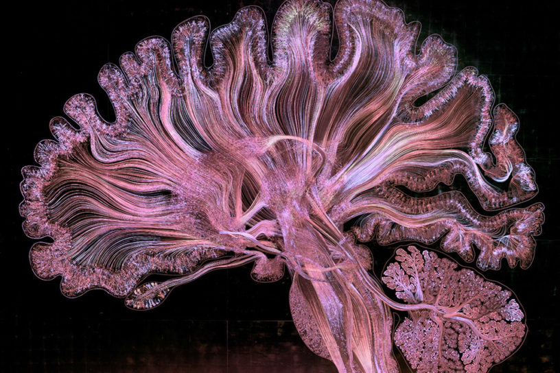
Taking nearly two years to complete, artist and neuroscientist Dr. Greg Dunn, along with his collaborator Dr. Brian Edwards, have mapped the neurons in the brain for a series of images titled Self Reflected. Produced through a technique they call reflective microetching, the two cross-disciplinary artists track the neural choreography in the mind, creating brilliant images that glow with a metallic luminescence.

The works depict a thin slice of the human brain at 22x the normal scale, each created through a combination of hand drawing, neuroscientific data, algorithmic simulation of neural circuitry, photolithography, strategic lighting design, and 1,750 sheets of 22k gold leaf.

“My work is neonaturalist, art based on natural forms and influenced by scientific advancements that allows us to perceive the universe beyond human senses,” explains Dunn in his artist statement. “Neonaturalism harmonizes unfamiliar scientific imagery and techniques with an experimental artistic scaffolding.”

Self Reflected was funded the National Science Foundation, and its first iteration is on permanent view at the Franklin Institute in Philadelphia, PA. Fine art prints and microetchings can be purchased on Dunn’s website. You can watch the work twinkle as it engages with a light source in the short video below. (via My Modern Met)

Self Reflected (detail), 22K gilded microetching, 96″ X 130″, 2014-2016, Greg Dunn and Brian Edwards. The brainstem and cerebellum, regions that control basic body and motor functions. (photo by Greg Dunn and Will Drinker)

Self Reflected (detail), 22K gilded microetching, 96″ X 130″, 2014-2016, Greg Dunn and Brian Edwards. The brainstem and cerebellum, regions that control basic body and motor functions. (photo by Greg Dunn and Will Drinker)

Self Reflected (detail), 22K gilded microetching, 96″ X 130″, 2014-2016, Greg Dunn and Brian Edwards. The thalamus and basal ganglia, sorting senses, initiating movement, and making decisions. (photo by Greg Dunn and Will Drinker)

Self Reflected (detail), 22K gilded microetching, 96″ X 130″, 2014-2016, Greg Dunn and Brian Edwards. The thalamus and basal ganglia, sorting senses, initiating movement, and making decisions. (photo by Greg Dunn and Will Drinker)

Self Reflected, 22K gilded microetching, 96″ X 130″, 2014-2016, Greg Dunn and Brian Edwards. The entire Self Reflected microetching under white light. (photo by Greg Dunn and Will Drinker)

Self Reflected, 22K gilded microetching, 96″ X 130″, 2014-2016, Greg Dunn and Brian Edwards. The entire Self Reflected microetching under white light. (photo by Greg Dunn and Will Drinker)

Self Reflected (detail), 22K gilded microetching, 96″ X 130″, 2014-2016, Greg Dunn and Brian Edwards. The visual cortex, the region located at the back of the brain that processes visual information.

Self Reflected (detail), 22K gilded microetching, 96″ X 130″, 2014-2016, Greg Dunn and Brian Edwards. The visual cortex, the region located at the back of the brain that processes visual information.

Self Reflected (detail), 22K gilded microetching, 96″ X 130″, 2014-2016, Greg Dunn and Brian Edwards. Raw colorized microetching data from the reticular formation.

Self Reflected (detail), 22K gilded microetching, 96″ X 130″, 2014-2016, Greg Dunn and Brian Edwards. Raw colorized microetching data from the reticular formation.

Self Reflected (detail), 22K gilded microetching, 96″ X 130″, 2014-2016, Greg Dunn and Brian Edwards. The pons, a region involved in movement and implicated in consciousness. (photo by Greg Dunn and Will Drinker)

Self Reflected (detail), 22K gilded microetching, 96″ X 130″, 2014-2016, Greg Dunn and Brian Edwards. The pons, a region involved in movement and implicated in consciousness. (photo by Greg Dunn and Will Drinker)

Self Reflected (detail), 22K gilded microetching, 96″ X 130″, 2014-2016, Greg Dunn and Brian Edwards. The parietal gyrus where movement and vision are integrated. (photo by Greg Dunn and Will Drinker)

Self Reflected (detail), 22K gilded microetching, 96″ X 130″, 2014-2016, Greg Dunn and Brian Edwards. The parietal gyrus where movement and vision are integrated. (photo by Greg Dunn and Will Drinker)

Self Reflected (detail), 22K gilded microetching, 96″ X 130″, 2014-2016, Greg Dunn and Brian Edwards. The motor and parietal cortex, regions involved in movement and sensation, respectively. (photo by Greg Dunn and Will Drinker)

Self Reflected (detail), 22K gilded microetching, 96″ X 130″, 2014-2016, Greg Dunn and Brian Edwards. The motor and parietal cortex, regions involved in movement and sensation, respectively. (photo by Greg Dunn and Will Drinker)

Self Reflected (detail), 22K gilded microetching, 96″ X 130″, 2014-2016, Greg Dunn and Brian Edwards. The midbrain, an area that carries out diverse functions in reward, eye movement, hearing, attention, and movement. (photo by Greg Dunn and Will Drinker)

Self Reflected (detail), 22K gilded microetching, 96″ X 130″, 2014-2016, Greg Dunn and Brian Edwards. The midbrain, an area that carries out diverse functions in reward, eye movement, hearing, attention, and movement. (photo by Greg Dunn and Will Drinker)

Self Reflected (detail), 22K gilded microetching, 96″ X 130″, 2014-2016, Greg Dunn and Brian Edwards. The laminar structure of the cerebellum, a region involved in movement and proprioception (calculating where your body is in space).

Self Reflected (detail), 22K gilded microetching, 96″ X 130″, 2014-2016, Greg Dunn and Brian Edwards. The laminar structure of the cerebellum, a region involved in movement and proprioception (calculating where your body is in space).

Related post

Play Sage Gateshead

Iconic Concert Hall becomes Play Sage Gateshead

Nima Rezai

Name: Nima Rezai. Hybridizing art, genres, technologies

Fink and AC3 IMMERSIVE ROOM

Fink and AC3 STUDIO for Who’s Next Paris Fair

[do_widget adsense_728x90_1]